Antibody Discovery
The diversity of the rabbit immune system yields picomolar-affinity antibodies that recognise hidden epitopes and small-molecule antigens.
The rabbit immune system's distinctive affinity-maturation machinery yields monoclonals of higher affinity and broader epitope coverage — particularly valuable for hard-to-detect targets such as small molecules, modified antigens and transmembrane proteins.
From immunization design through positive-clone screening, evaluation is function-driven and keyed to the intended use — neutralisation, blocking, IHC or flow cytometry.
Development is customised across antigen classes — weak immunogens, transmembrane proteins, modified antigens — for immunoassay, protein-interaction and clinical-research applications.
Enquire now →

The rabbit immune system's distinctive affinity-maturation machinery yields monoclonals of higher affinity and broader epitope coverage — particularly valuable for hard-to-detect targets such as small molecules, modified antigens and transmembrane proteins.
From immunization design through positive-clone screening, evaluation stays keyed to the antibody's real application — neutralisation, blocking, IHC, flow cytometry — so what is screened out is genuinely usable.
Immunization workflow, screening criteria and expression system are customised to the target and downstream use, locking critical quality attributes before kickoff and avoiding rework later.
Immunogen design, animal strategy and validation path are planned systematically at project inception, heading off non-specific binding and loss of function and materially shortening development time.

Eight stages. You may supply the antigen (peptide purity ≥95%, protein purity ≥85%); outputs and acceptance criteria for each stage are listed below.
Immunization plan customised to the target and downstream use; antigen supplied or commissioned
The antigen is prepared and checked for purity, concentration and integrity
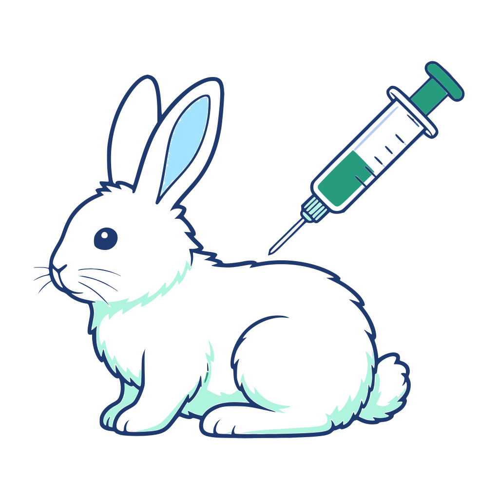
8–10 weeks of immunization; titre after the 3rd–4th boost ≥1:256,000
Titre and specificity results are reviewed before screening begins
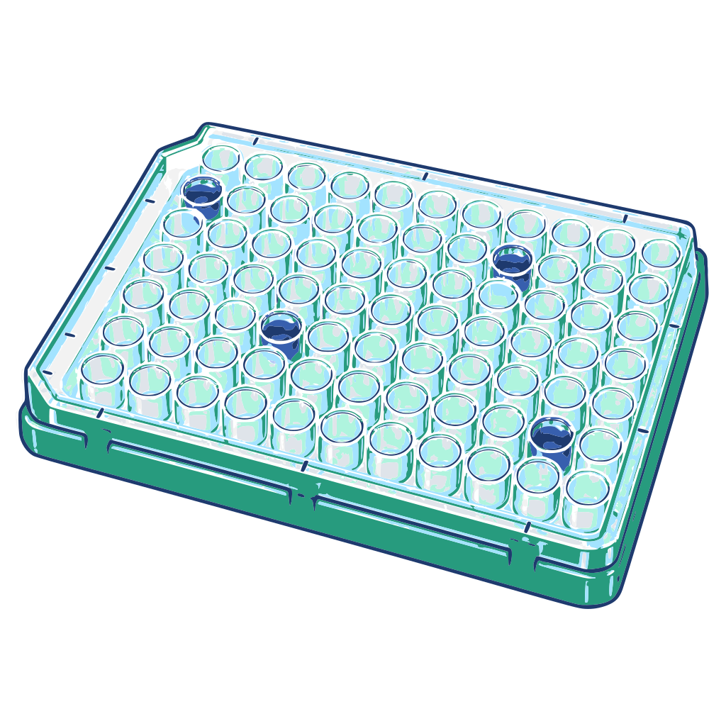
Screened by single B cell / phage display; ELISA-positive
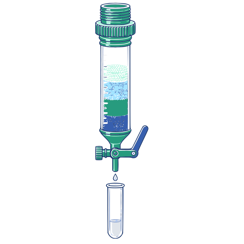
Transient Expi293 expression and purification — ≥1 mg at ≥95% purity
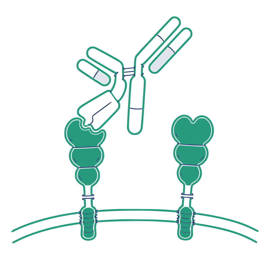
Titre, affinity and specificity verified by ELISA/WB, EC50 <10 nM
Purified antibody, sequence data and binding-curve CoA delivered
Immunization through QC totals roughly 16–24 weeks; commissioned antigen preparation adds 3–5 weeks.
From immunization design to positive-clone screening, evaluation is oriented around the real application — neutralisation, blocking, IHC, flow cytometry. Critical quality attributes are locked before kickoff, avoiding rework later.
Deliverables, acceptance criteria and timelines by experimental stage. The staging differs from the process above: the process shows how a project runs, while this table is the acceptance basis written into the contract.
| Stage | Scope | Deliverables | Standards | Timeline |
|---|---|---|---|---|
| Antigen preparation | Supplied by you or prepared by SanjingBio | Peptide / protein | Peptide purity ≥95%, protein purity ≥85% | 3–5 weeks |
| Animal immunization | Rabbit immunization | Immunization record | / | 8–10 weeks |
| Serum titre testing | Serum titre after the 3rd–4th boost | Titre results | Titre ≥1:256,000 | 2–3 days |
| Antibody screening | Single B cell / phage display | Positive clones | ELISA-positive | 4–8 weeks |
| Gene retrieval & plasmid construction | Sequencing and vector construction | / | Sequence | 2–3 weeks |
| Transient expression and purification | Expi293 expression and purification | ≥1 mg purified antibody | Purity ≥95% | 1–2 weeks |
| QC testing | ELISA / WB validation | Antibody binding-curve CoA report | EC50 <10 nM | 1 week |
Using the rabbit hybridoma route, immunization with recombinant target protein plus eukaryotic expression cells — followed by multiple rounds of subcloning and high-throughput screening — produced target monoclonals of high specificity and sensitivity that perform in both IF and FCM.
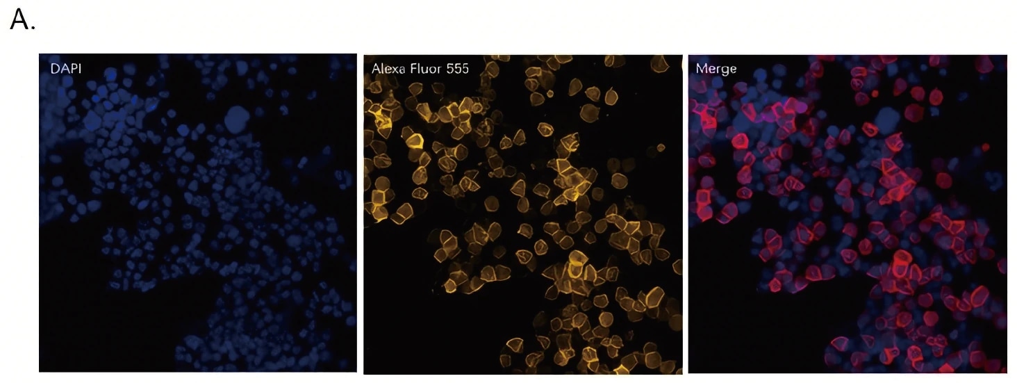
Co-localisation of nuclei (blue), the target protein (yellow) and antibody binding signal (red).
Panels from left to right: DAPI, Alexa Fluor 555, merge.
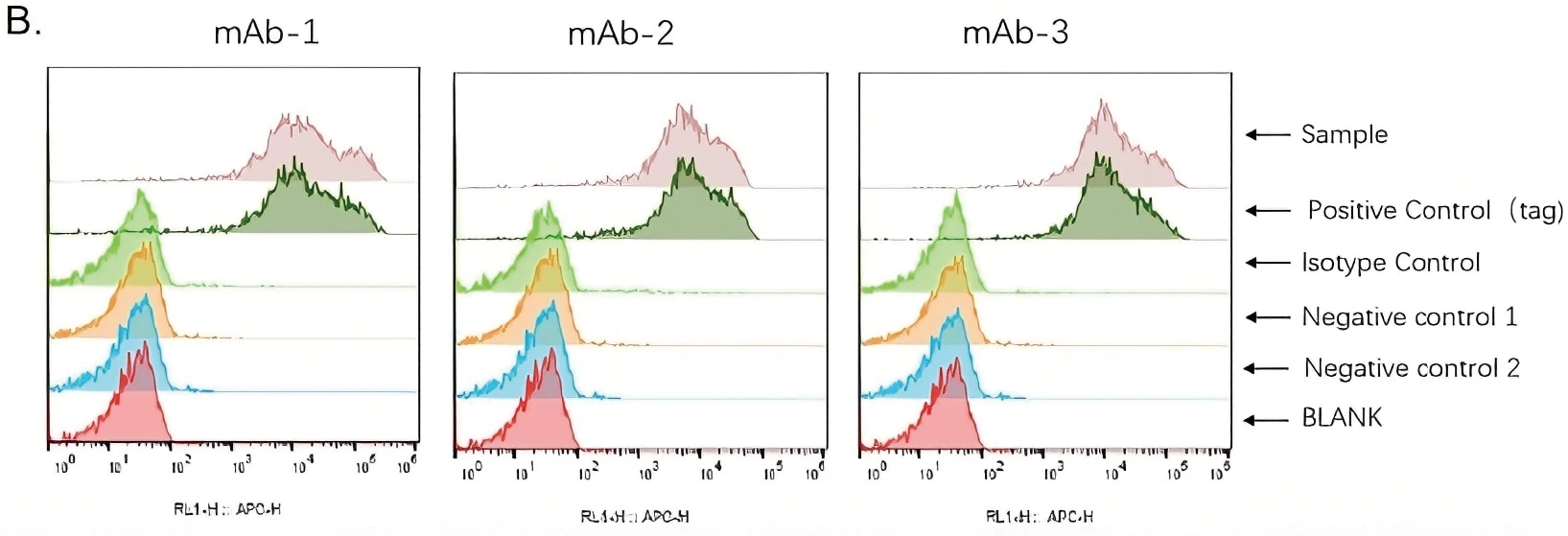
Binding levels of the monoclonals across samples, referenced against isotype control, negative controls and blank.
Panels: mAb-1 / mAb-2 / mAb-3. Within each panel the six traces top to bottom are Sample, Positive Control (tag), Isotype Control, Negative control 1, Negative control 2 and BLANK; x-axis RL1-H :: APC-H.
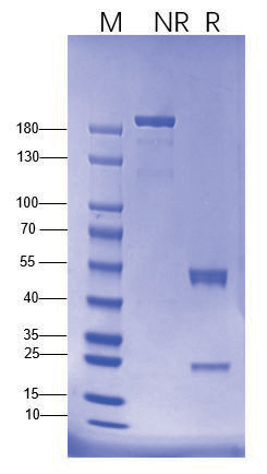
Electrophoresis under reducing/non-reducing (R/NR) conditions shows high antibody purity, clean subunit composition, correct disulfide state and an intact overall structure with impurities under control.
Share the intended application and what you already have in hand. A scientist — not a sales rep — will scope feasibility, suggest the right route and send a quote, usually within one business day.