

May 27, 2026
Breast cancer remains one of the most prevalent malignancies among women, posing a substantial threat to physical and mental health. Among all breast cancer cases, approximately 15%–20% are classified as the "HER2-positive" subtype, which is characterized by more aggressive proliferation and invasive potential, and was historically associated with poor prognosis. However, advances in understanding HER2 as a therapeutic target and the subsequent development of targeted agents have substantially improved clinical outcomes for patients with HER2-positive breast cancer.
HER2 (human epidermal growth factor receptor 2) is a transmembrane glycoprotein localized on the cell surface, belonging to the epidermal growth factor receptor (EGFR/ErbB) family. Its primary function is to serve as a ligand-dependent signal-transducing receptor that mediates the activation of signaling pathways involved in cell proliferation, differentiation, and survival. Under physiological conditions, the HER2 gene is located on chromosome 17q21 with a copy number of 2, and its expression is tightly regulated, with low-level expression detectable only in a limited range of normal epithelial tissues. In a subset of breast cancers, however, HER2 gene amplification and/or transcriptional upregulation leads to overexpression of the encoded protein on the cell membrane, defined as the "HER2-positive" status. This pathological alteration markedly enhances the activation intensity of downstream pro-proliferative signaling cascades, including the PI3K/AKT and MAPK pathways, conferring a selective growth advantage to tumor cells and correlating closely with unfavorable clinical prognosis.
The value of HER2 as a therapeutic target is rooted in its driving role in tumor initiation and progression. HER2-positive breast cancer thus constitutes an independent molecular subtype that exhibits high sensitivity to HER2-directed therapies, such as trastuzumab, pertuzumab, and antibody-drug conjugates. Accurate determination of HER2 status in clinical practice relies on standardized pathological testing methods, including immunohistochemistry (IHC) for semi-quantitative assessment of protein expression levels, and fluorescence in situ hybridization (FISH) for quantitative analysis of gene copy numbers. These test results not only serve as the gold standard for determining HER2 status but also represent the core basis for formulating individualized treatment strategies. HER2 testing therefore plays a pivotal "biomarker-navigation" role in the precision medicine framework for breast cancer, directly impacting the screening of indications for targeted therapy and the prediction of therapeutic efficacy.
The clinical implementation of HER2-targeted therapy was revolutionized by the development and regulatory approval of trastuzumab, a recombinant humanized IgG1 monoclonal antibody. Trastuzumab binds with high affinity to domain IV of the extracellular region of the HER2 receptor and exerts its anti-tumor effects through a dual mechanism: directly inhibiting HER2-mediated downstream signal transduction, and simultaneously mobilizing the innate immune system to eliminate tumor cells via antibody-dependent cell-mediated cytotoxicity (ADCC). The introduction of trastuzumab fundamentally altered the natural history of HER2-positive breast cancer, significantly reducing the risk of recurrence in early-stage patients and extending overall survival in those with metastatic disease.
Building on the success of trastuzumab, researchers further explored the feasibility of combination blockade strategies. Pertuzumab, another humanized monoclonal antibody, binds to a distinct epitope located in domain II of the HER2 extracellular region, which corresponds to the dimerization domain. This binding site is entirely different from that of trastuzumab, and the two antibodies do not compete with each other spatially. Instead, they act synergistically to more comprehensively block heterodimerization of HER2 with other HER family members (such as HER3), thereby achieving more thorough suppression of downstream pro-proliferative signals. Clinical studies have demonstrated that the dual-targeted combination of trastuzumab and pertuzumab, when used in neoadjuvant and adjuvant settings for HER2-positive early breast cancer, significantly improves pathological complete response rates and long-term disease-free survival compared with single-agent regimens. The 2025 edition of the Chinese Expert Consensus on Targeted HER2 Breast Cancer Diagnosis and Treatment has recommended this combination as a standard-of-care treatment for patients at high risk of recurrence.
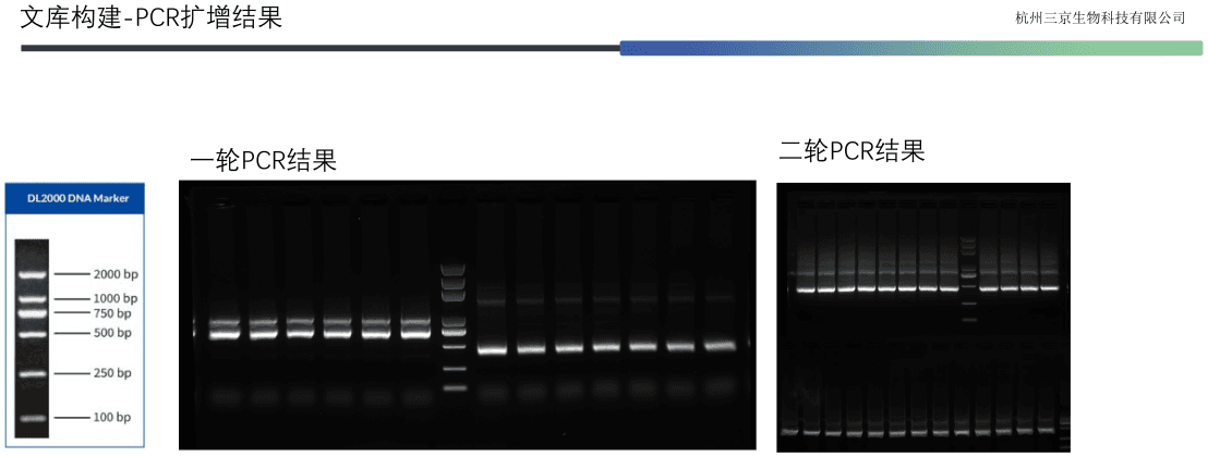
The discovery of targeted therapeutic agents depends critically on high-quality bioactive materials and robust screening systems. In early-stage research and development, the preparation of antigenic proteins with native conformation and favorable immunogenicity is a prerequisite for obtaining functional antibodies. Experimental records from Hangzhou Sanjing Biotechnology Co., Ltd. (data as of June 17, 2026) indicate that researchers employed HER2-His recombinant protein (SJR199, UniProt accession P04626, amino acid residues Thr23-Ser635) as the immunogen. This protein encompasses the extracellular domain of the HER2 receptor and is capable of mimicking its native spatial conformation on the cell surface. ELISA validation confirmed that this immunogen could be specifically recognized by a commercially available anti-HER2 monoclonal antibody (Acro, HE2-M598), demonstrating satisfactory antigenicity (Figure 1).
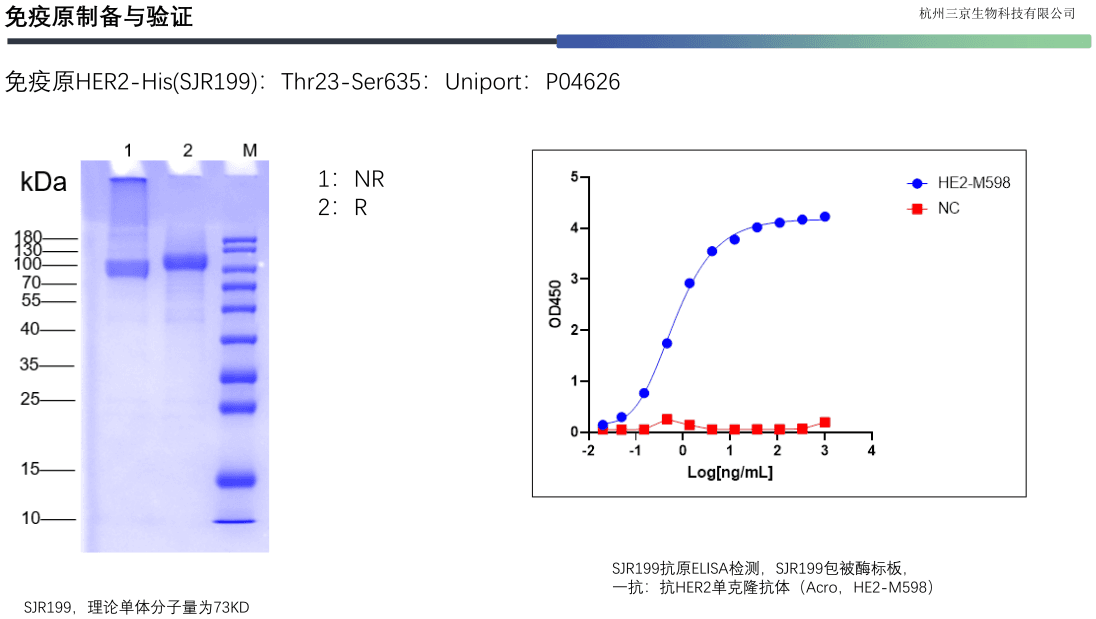
To obtain nanobodies (VHH) with unique binding epitopes—a class of heavy-chain single-domain antibodies derived from camelids—the researchers selected alpacas as the immunization host. The magnitude of the immune response was quantitatively assessed by serum titer (Figure 2). The data showed that after the third booster immunization, the VHH serum titer against HER2 reached 4,420 (calculated using OD450 = 0.2 as the cutoff value), and the IgG titer reached 288,956, both far exceeding the conventional library construction threshold (VHH titer > 1,000). These results confirmed that the immunogen successfully elicited a robust humoral immune response in the host, providing a solid foundation for the subsequent construction of a large, highly diverse nanobody gene library.
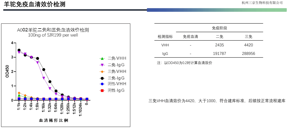
Following confirmation of the immune response, the experiment proceeded to the isolation and amplification of antibody genes. Figure 3 presents the agarose gel electrophoresis results of the first-round polymerase chain reaction (PCR). This step was designed to specifically amplify gene fragments encoding the nanobody variable region from the total mRNA of peripheral blood lymphocytes of immunized alpacas. The clear target bands on the gel indicated successful amplification of high-quality VHH gene products, which are the core raw material for the construction of phage display or yeast display nanobody libraries.
Candidate antibody molecules selected from gene libraries must undergo functional validation at the cellular level to evaluate their binding capacity to native conformation antigens and their species compatibility. In preclinical drug development, candidate molecules that exhibit cross-reactivity with both human and monkey antigens are particularly valuable, as they enable toxicological and pharmacokinetic evaluation in non-human primate models.
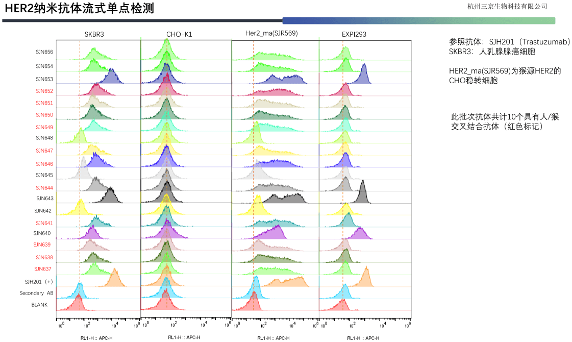
Figure 4 shows the results of flow cytometry (FCM)-based detection. The researchers simultaneously used the human breast cancer cell line SKBR3 (endogenously overexpressing HER2) and a CHO engineered cell line stably transfected with monkey HER2 (HER2_Ma) as target cells to screen a panel of candidate nanobodies for binding activity. Flow cytometry enables quantitative detection of the binding efficiency of fluorescently labeled antibodies to cell surface target proteins at single-cell resolution. The results showed that a total of 10 molecules (marked in red in the figure) from this batch of candidate antibodies exhibited cross-reactivity, binding to both human and monkey HER2. SJH201 (i.e., trastuzumab) was used as a positive reference antibody in the figure to calibrate the experimental system and compare relative binding intensities.
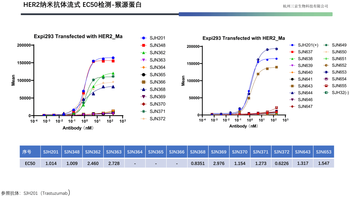
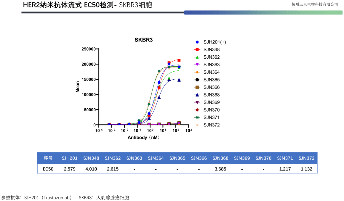
To further quantify the binding affinity of candidate molecules, Figures 5 and 6 present the half-maximal effective concentration (EC50) data for two batches of candidate antibodies. The EC50 value, as a critical parameter for assessing antibody-antigen binding efficiency, is inversely correlated with affinity. Using trastuzumab (SJH201) as the reference standard, the tabulated data list the EC50 values for early-stage candidate molecules (SJH348, SJH362, etc.) in Figure 5, measured on monkey HER2-expressing cells, and for subsequent candidate molecules (SJH643, SJH653, etc.) in Figure 6, measured on SKBR3-expressing cells. These quantitative data provide direct scientific evidence for ranking the affinity of different molecules, selecting lead compounds, and guiding subsequent affinity maturation efforts.
The HER2-targeted therapy field is currently evolving toward greater diversification and precision. The following directions merit particular attention:
Clinical application of antibody-drug conjugates (ADCs): ADC agents such as trastuzumab emtansine (T-DM1) and trastuzumab deruxtecan (T-DXd) conjugate HER2-targeting monoclonal antibodies with highly cytotoxic small-molecule payloads via cleavable or non-cleavable linkers. This design combines the targeting delivery capability of antibodies with the potent killing activity of cytotoxic agents, enabling precise elimination of antigen-positive tumor cells. For HER2-positive breast cancer patients who do not achieve pathological complete response after neoadjuvant therapy, T-DM1 is currently the standard recommended adjuvant treatment option.
Optimization of administration modalities: Traditional dual-targeted intravenous infusion is time-consuming. The development of Phesgo®, a novel fixed-dose subcutaneous formulation containing both trastuzumab and pertuzumab, has reduced administration time to 5–8 minutes, significantly improving treatment convenience and patient compliance while optimizing the allocation of medical resources.
Individualized stratification of treatment intensity: Not all HER2-positive patients require the maximum-intensity combination therapy. For low-risk patients with low tumor burden and node-negative status, an appropriate "de-escalation" approach can reduce unnecessary toxicity while maintaining efficacy. In the future, dynamic risk stratification using multigene testing tools (such as HER2DX) is expected to enable more precise matching of treatment intensity.
The therapeutic development journey for HER2-positive breast cancer exemplifies the successful translation of fundamental scientific discoveries into clinical practice. From elucidation of the biological function of the HER2 gene, to the development of monoclonal antibodies, and through continued iterative innovation in dual-targeted combinations, ADCs, and formulation optimization, each advance has been built upon rigorous laboratory research, systematic preclinical evaluation, and well-designed prospective clinical trials. This trajectory has not only profoundly improved patient survival outcomes but has also provided a replicable paradigm for the development of targeted therapies in solid tumors. Continued innovation in this field will remain a major driving force in the advancement of precision oncology.


| Product Name | Catalog No. | Product Name | Catalog No. |
|---|---|---|---|
| Oncology | Oncology | ||
| Cadherin-17 (CDH17) | SJ0501 | Ectonucleoside triphosphate diphosphohydrolase-1 (CD39) | SJ6901 |
| Carcinoembryonic antigen-related cell adhesion molecule 5 (CEACAM5) | SJ0601 | 5'-nucleotidase (CD73) | SJ7001 |
| Mesothelin (MSLN) | SJ0801 | Glypican-3 (GPC3) | SJ7101 |
| Protein tyrosine kinase 7 (PTK7) | SJ1001 | Insulin-like growth factor 1 receptor (1GF-1R) | SJ7201 |
| 5T4 oncofetal antigen (5T4) | SJ1101 | C-C chemokine receptor 2 (CCR2) | SJ7401 |
| Guanylate cyclase C (GUCY2C) | SJ1201 | C-C chemokine receptor 7 (CCR7) | SJ7501 |
| Immunoregulatory checkpoint glycoprotein (B7H3) | SJ1301 | C-C chemokine receptor 9 (CCR9) | SJ7601 |
| Human epidermal growth factor receptor 2 (HER2) | SJ1401 | Folate receptor alpha (FRα) | SJ0301 |
| Human epidermal growth factor receptor 3 (HER3) | SJ1701 | Carcinoembryonic antigen-related cell adhesion molecule 6 (CEACAM6) | SJ0701 |
| Adenocarcinoma-associated antigen (EPCAM) | SJ1801 | Seizure-related protein 6 (SEZ6) | SJ6701 |
| Hepatocyte growth factor receptor (MET) | SJ1901 | Podoplanin (PDPN) | SJ7701 |
| Interleukin-3 receptor alpha chain (IL3RA) | SJ2001 | Cadherin-6 (CDH6) | SJ8101 |
| Leukocyte differentiation antigen (CD19) | SJ2201 | Protocadherin beta-9 (PCDHB9) | SJ8201 |
| T-cell antigen CD7 (CD7) | SJ2301 | Fibroblast growth factor receptor 4 (FGFR4) | SJ8301 |
| B-cell maturation antigen (BCMA) | SJ2401 | Integrin alpha-2 (ITGA2) | SJ8401 |
| Programmed death-ligand 1 (PDL1) | SJ2501 | Melanoma cell adhesion molecule (MCAM) | SJ8501 |
| Six-transmembrane epithelial antigen of the prostate 1 (STEAP1) | SJ2601 | LY6/PLAUR domain-containing protein 3 (LYPD3) | SJ8801 |
| Kallikrein-2 (KLK2) | SJ2701 | Frizzled-2 (FZD2) | SJ8901 |
| TNF-like ligand 1A (TL1A) | SJ2801 | Frizzled-6 (FZD6) | SJ9001 |
| Programmed death-ligand 1 (PDL1) | SJ3001 | Frizzled-8 (FZD8) | SJ9101 |
| C-C chemokine receptor 6 (CCR6) | SJ3101 | CD226 molecule (CD226) | SJ9201 |
| B-cell antigen receptor complex-associated protein beta chain (CD79B) | SJ3201 | Cellular communication network factor 4 (CCN4) | SJ9301 |
| Fibroblast activation protein (FAP) | SJ3301 | Collagen triple helix repeat-containing protein 1 (CTHRC1) | SJ9401 |
| Integrin beta-1 (ITGB1) | SJ3401 | Fibronectin-1 (FN1) | SJ9501 |
| Leucine-rich repeat-containing G-protein-coupled receptor 5 (LGR5) | SJ3501 | Discoidin domain receptor tyrosine kinase 2 (DDR2) | SJ9601 |
| Leucine-rich repeat-containing G-protein-coupled receptor 6 (LGR6) | SJ3601 | Vascular endothelial growth factor A (VEGFA) | SJ9701 |
| Receptor-type tyrosine-protein phosphatase H (PTPRH) | SJ3701 | C-X-C motif chemokine ligand 12 (CXCL12) | SJ9801 |
| Solute carrier family 2 member 1 (SLC2A1) | SJ3801 | Transforming growth factor beta 1 (TGFB1) | SJ9901 |
| Sphingosine-1-phosphate receptor 5 (S1PR5) | SJ4001 | Autoimmune | |
| Tumor-associated calcium signal transducer 2 (TACSTD2) | SJ4101 | Ephrin-A5 (EFNA5) | SJ2101 |
| Poliovirus receptor-related protein 4 (Nectin-4) | SJ4201 | Leukocyte differentiation antigen (I1L4R) | SJ2901 |
| Secreted phosphoprotein 1 (SPP1) | SJ4301 | Interleukin-20 receptor subunit beta (IL20RB) | SJ3901 |
| C-X-C chemokine receptor (CXCR5) | SJ4401 | Interleukin-4 (IL4) | SJ5301 |
| C-X-C chemokine receptor (CXCR6) | SJ4501 | Interleukin-11 (1L11) | SJ5901 |
| C-X-C chemokine receptor (CXCR3) | SJ4601 | Interleukin-33 (IL33) | SJ6401 |
| Sialic acid-binding immunoglobulin-like lectin 8 (Siglec8) | SJ4701 | Interleukin-15 (IL15) | SJ6501 |
| Sialic acid-binding immunoglobulin-like lectin 6 (Siglec6) | SJ4801 | Interleukin-6 signal transducer (IL6ST) | SJ7301 |
| T-lymphocyte antigen CD2 (CD2) | SJ4901 | Platelet endothelial aggregation receptor 1 (PEAR1) | SJ7901 |
| B-lymphocyte antigen CD20 (CD20) | SJ5201 | Metabolic | |
| Claudin-1 (CLDN1) | SJ5401 | Activin receptor type IIA (ActRIIA) | SJ1501 |
| T-cell surface glycoprotein CD8 alpha chain (CD8A) | SJ5501 | Transferrin receptor 1 (TFR1) | SJ1601 |
| T-cell surface glycoprotein CD8 beta chain (CD8B) | SJ5601 | Integrin beta-8 (ITGB8) | SJ5001 |
| Leukocyte antigen CD37 (CD37) | SJ5701 | Glucose-dependent insulinotropic polypeptide receptor (GIPR) | SJ5101 |
| Leucine-rich repeat-containing protein 15 (LRRC15) | SJ6001 | Cadherin-3 (CDH3) | SJ5801 |
| Delta-like ligand 3 (DLL3) | SJ6101 | Solute carrier family 1 member 5 (SLC1A5) | SJ8601 |
| Delta-like ligand 4 (DLL4) | SJ6201 | Solute carrier family 22 member 7 (SLC22A7) | SJ8701 |
| Prostate-specific membrane antigen (PSMA) | SJ6301 | Other | |
| Zinc transporter LIV-1 (LIV-1) | SJ6601 | Lysophosphatidic acid receptor 1 (LPAR1) | SJ7801 |
| Interleukin-17A (IL17A) | SJ6801 | Tissue factor (FIII) | SJ8001 |
May 27, 2026
Jul 15, 2026

Jun 10, 2026
Share the intended application and what you already have in hand. A scientist — not a sales rep — will scope feasibility, suggest the right route and send a quote, usually within one business day.